
Vitamin A is another hormone gaining popularity recently as more people research nutrition.
Hormones function differently than vitamins or minerals. To support hormones, we need to support a handful of nutrients and metabolic processes. That way the hormone has a clear line to function in.
Hormones are similar to gauges on the dashboard of our vehicle. The gasoline an engine runs on is like a nutrient, and the temperature gauge reading is like a hormone level. If the hormone level is incorrect, the hormone itself has very little to do with the issue.
An issue with Vitamin A is an issue with things like sulfite and ceruloplasmin metabolism, which each depend on a handful of various nutrients and proper bile function etc.
The molybdenum cofactor (MoCo) serves as the essential prosthetic group for sulfite oxidase, the terminal enzyme converting toxic sulfite to sulfate in cysteine metabolism[1]. When MoCo synthesis fails or molybdenum becomes deficient, sulfite accumulates and depletes cellular glutathione through formation of glutathione S-sulfonate (GSSO3H), a competitive inhibitor of glutathione S-transferases with Ki values of 4-14 μM[2]. This glutathione depletion cascade directly impairs the sulfation pathways required for vitamin A detoxification.
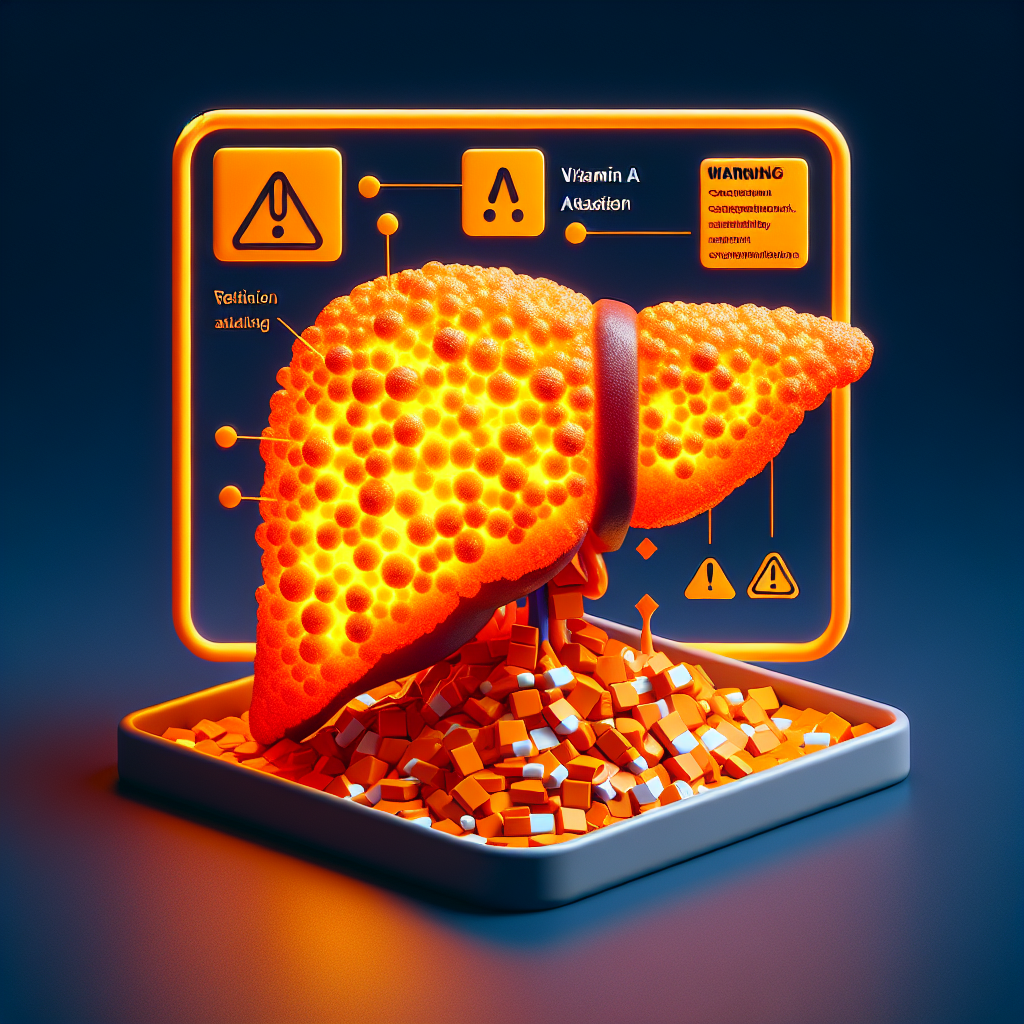
When I refer to vitamin A “detoxification,” I’m describing the essential process of conjugating retinol and its metabolites for cellular export and elimination. This isn’t about vitamin A being a toxin per se, but rather about the body’s need to precisely regulate cellular retinol levels through controlled elimination pathways. When these pathways fail, vitamin A accumulates intracellularly, creating toxicity through the very mechanisms meant to be beneficial.
Research demonstrates that vitamin A metabolism depends heavily on Phase II sulfation for cellular detoxification. All-trans retinoic acid directly induces multiple sulfotransferase enzymes – SULT1A1, SULT2A1, and SULT1E1 – at the transcriptional level, with intestinal sulfotransferases showing greater responsiveness than hepatic enzymes[3]. The obligate cosubstrate for these reactions, PAPS (3′-phosphoadenosine-5′-phosphosulfate), can be depleted within 2 minutes during active sulfation[4]. When sulfite oxidase dysfunction prevents normal sulfite-to-sulfate conversion, PAPS synthesis becomes compromised, creating a bottleneck that prevents proper vitamin A conjugation and elimination.
The connection extends through bile acid metabolism, which research from 2019 reveals as critical for vitamin A homeostasis. The Farnesoid X Receptor (FXR) heavily regulates hepatic vitamin A storage, with FXR-null mice showing over 90% reduction in hepatic retinol and retinyl palmitate levels[5]. Molybdenum deficiency and sulfite accumulation disrupt bile acid synthesis, further compromising vitamin A absorption and storage capacity.
Cascading trace mineral disruptions amplify vitamin A toxicity
Sulfite-induced glutathione depletion triggers a devastating cascade affecting multiple trace mineral-dependent systems essential for vitamin A metabolism. The selenium-dependent glutathione peroxidase system, which requires adequate glutathione as its reducing substrate, becomes functionally impaired even when selenium levels remain normal[6]. This creates synergistic oxidative stress that overwhelms cellular detoxification capacity.
Ceruloplasmin, the copper-containing ferroxidase carrying over 95% of plasma copper, demonstrates remarkable vitamin A dependency[7]. Retinoic acid injection increases ceruloplasmin activity up to 4-fold within 4 days through direct transcriptional regulation. However, when sulfite metabolism fails and oxidative stress increases, ceruloplasmin synthesis becomes impaired, creating a vicious cycle where vitamin A cannot properly regulate its own transport proteins.
Zinc’s role proves equally critical – zinc deficiency significantly impairs hepatic synthesis of retinol-binding protein (RBP), the specific transport protein for vitamin A[8]. Studies in HepG2 cells reveal a paradoxical 7.5-fold increase in RBP mRNA during zinc deficiency as cells attempt compensatory upregulation, yet actual RBP secretion and vitamin A mobilization remain blocked[9]. The zinc-dependent alcohol dehydrogenases required for retinol-to-retinal conversion also fail, trapping vitamin A in unusable forms[10].
Individual genetic variations determine toxicity thresholds
The remarkable variability in vitamin A tolerance – ranging from toxicity at 25,000 IU daily in some individuals to others tolerating much higher doses – stems from multiple genetic factors interacting with sulfur metabolism status[11]. Up to 45% of the population carries BCMO1 gene variants (rs7501331 and rs12934922) that reduce beta-carotene conversion efficiency by up to 69%[12]. These “poor converters” may compensate with enhanced absorption mechanisms that predispose them to toxicity when consuming preformed vitamin A.
Isolated sulfite oxidase deficiency, caused by SUOX gene mutations, presents the clearest example of sulfur metabolism disrupting vitamin A handling[13]. Patients accumulate toxic levels of sulfite, S-sulfocysteine, and thiosulfate while experiencing decreased cysteine availability[14]. The nervous system proves particularly vulnerable, with classical presentations including neonatal seizures, feeding difficulties, and progressive neurological deterioration[15]. Three subtypes of molybdenum cofactor deficiency (MoCD) produce similar metabolic disruptions, with Type A now treatable with fosdenopterin (synthetic cyclic pyranopterin monophosphate)[16].
Historical research from 1965 using radioactive sulfur tracers demonstrated that vitamin A overdose causes increased protein breakdown and altered sulfate metabolism, with more rapid decrease in urinary sulfate specific activity[17]. This proteolytic effect releases additional sulfur-containing amino acids, potentially overwhelming already compromised sulfite oxidase capacity.
Phase II detoxification bottlenecks trap vitamin A in cells
The primary mechanism for vitamin A elimination involves coordinated Phase I hydroxylation by CYP26 enzymes (CYP26A1, CYP26B1, CYP26C1) producing 4-OH-retinoic acid and 4-oxo-retinoic acid, followed by Phase II conjugation[18]. UGT2B7 serves as the only human UDP-glucuronosyltransferase capable of glucuronidating retinoids, forming retinyl β-glucuronide and retinoyl β-glucuronide found at mean serum concentrations of 6.8 ± 4.0 nmol/L and 2.42 ng/mL respectively[19].
When sulfation pathways fail due to PAPS depletion or sulfotransferase dysfunction, the entire burden shifts to UGT2B7-mediated glucuronidation[20]. However, pharmacological retinoid concentrations cause rapid UGT2B7 down-regulation in intestinal cells, creating a metabolic trap where vitamin A cannot be properly conjugated for elimination[21]. This explains why some individuals quickly develop toxicity symptoms – their compromised sulfation capacity combined with overwhelmed glucuronidation prevents cellular vitamin A export.
Research reveals tissue-specific responses, with intestinal tissues showing greater sulfotransferase induction by retinoic acid compared to hepatic tissues[22]. This differential regulation suggests that individuals with intestinal dysfunction may be particularly vulnerable to vitamin A accumulation when sulfur metabolism becomes impaired.
Clinical patterns reveal metabolic signatures of intolerance
Clinical observations demonstrate clear patterns linking sulfur metabolism disruption to vitamin A intolerance. Patients with compromised liver function – whether from alcohol consumption, viral hepatitis, or protein-energy malnutrition – show toxicity at doses as low as 25,000 IU daily, compared to healthy individuals tolerating higher amounts[23]. Water-miscible vitamin A preparations prove more toxic than oil-based forms, likely due to rapid absorption overwhelming compromised detoxification systems[24].
The therapeutic window narrows dramatically in metabolically compromised individuals. While Arctic populations historically consumed polar bear liver containing 12-26,000 IU/gram with acute toxicity[25], some modern patients develop advanced hepatic fibrosis from 220,000 IU daily supplementation[26]. Children prove particularly sensitive, with toxicity reported at 1,500 IU/kg body weight[27].
Molybdenum cofactor deficiency patients require comprehensive metabolic support beyond simple supplementation. Type A MoCD responds to fosdenopterin treatment, which provides the synthetic cyclic pyranopterin monophosphate precursor needed for MoCo synthesis[28]. However, secondary thiamine deficiency occurs due to sulfite inactivation, and hyperlactatemia commonly develops from mitochondrial dysfunction[29].
Conclusion
The intricate relationship between vitamin A metabolism and sulfur/molybdenum pathways explains the dramatic individual variability in vitamin A tolerance. When sulfite oxidase function fails – whether through molybdenum deficiency, genetic mutations, or metabolic overwhelm – a cascade of disruptions prevents proper vitamin A detoxification and cellular export. The resulting accumulation triggers oxidative stress, depletes glutathione, impairs trace mineral-dependent proteins, and creates a self-perpetuating cycle of metabolic dysfunction. Understanding these interconnected pathways reveals why single-nutrient supplementation often fails and highlights the need for comprehensive metabolic assessment before vitamin A supplementation, particularly in individuals with signs of sulfur metabolism disruption.